We are excited to introduce you to a groundbreaking research study recently conducted using our AMBER X cryo-electron microscope. The scientific team from the University of Pennsylvania harnessed our technology to achieve previously unimaginable results in the field of neuroscience.
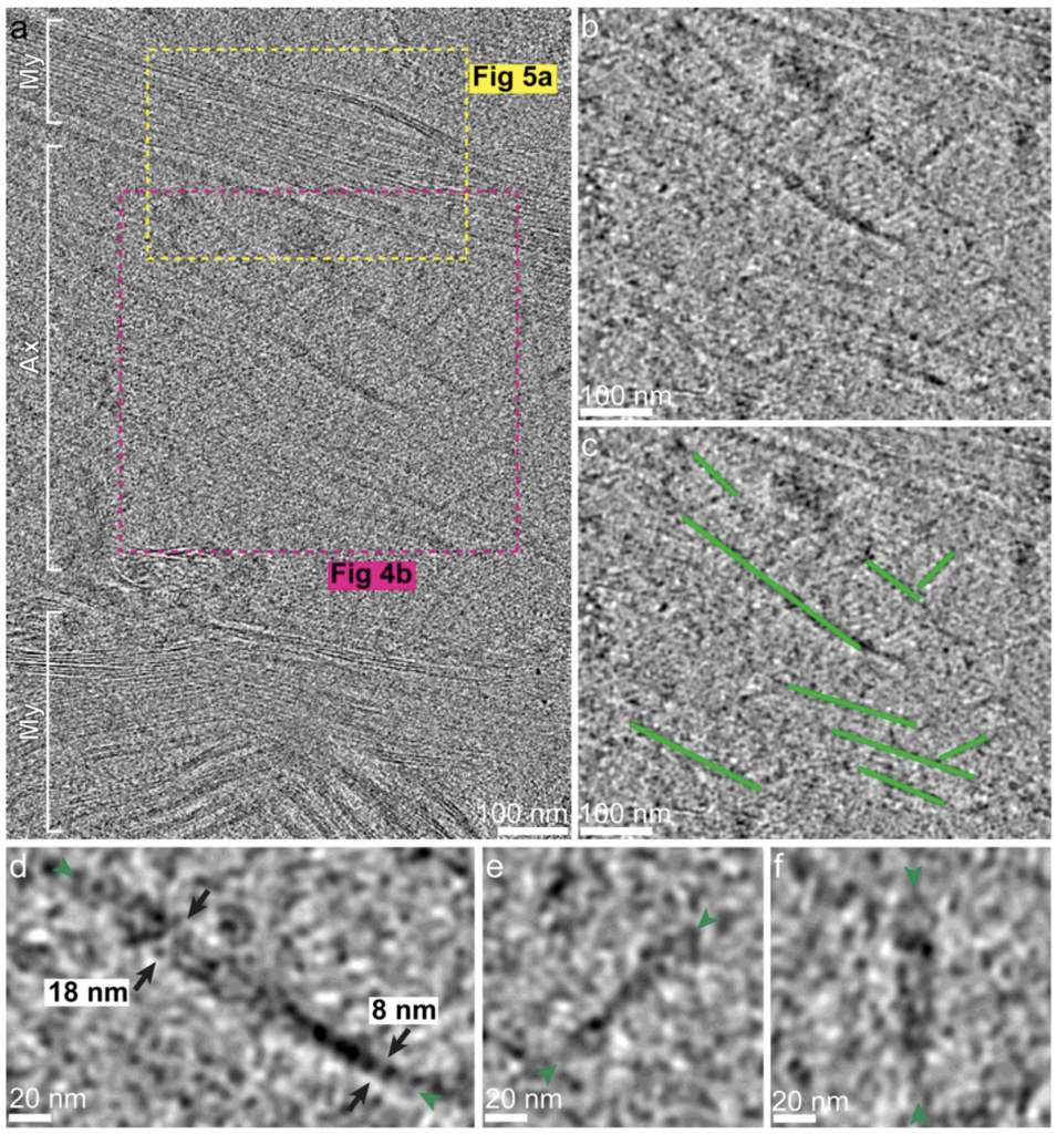
This team of experts obtained a sample of human brain tissue directly from an autopsy, which they then subjected to rapid freezing and cryo-FIB milling. Thanks to our microscope, the scientists were able to visualize intricate subcellular structures, such as myelin sheaths and tau aggregates, in their natural state and at an incredible resolution.
What does this mean for science? This marks the first instance where human brain tissue has been imaged with such precision and depth of detail. This breakthrough would not have been possible without the capabilities of our microscope, underscoring its significance in modern research. We are immensely proud that our technology could contribute to such a monumental advancement. The ability to examine human tissue at the subcellular level may open new avenues in understanding neurological diseases.
This is a significant achievement by the R&D FIB-SEM Applications team led by Rostislav Váňa, specifically Samuel Záchej and Martina Zánová. It’s a testament to the quality of our technology and the expertise of our staff that researchers choose our instrument and rely on our support for conducting such groundbreaking studies.
Stories like this fuel our daily passion and spark inspiration for our next innovations. We extend our gratitude to all colleagues across the company who contribute to the success of TESCAN devices. Together, we continue to push the boundaries of what’s possible in science and technology.